
Figure 3.
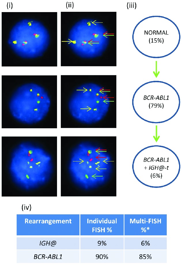
Images of FISH interphase nuclei depicting the apparent emergence of a BCR-ABL1 translocation prior to the IGH@-t. (i) Captured interphase nuclei from patient 21733 hybridized with FISH probes to track BCR-ABL1 (DAKO BCR split signal probe; red/green) and a home-made IGH@ probe (gold). (ii) The same captured inter-phase nuclei, including colored arrows highlighting the signals that were scored: the yellow arrows highlight the gold probe marking the home-made IGH@ probe that detects both the copy number of chromosome 14 and also the presence of a translocation. The red/green double arrows highlight the BCR split signal probe that marks the BCR-ABL1 translocation. (iii) Cartoon depicting a possible model of evolutionary progression by gain (green arrows). A normal population of cells was observed (15%) with the emergence of a clone harboring only the BCR-ABL1 translocation in 79% of nuclei. This clone subsequently acquired an IGH@-t in 6%. (iv) Table showing the percentages of positive nuclei when samples were hybridized with either the individual probes for each rearrangement (Individual FISH), or when they were investigated together (Multi-FISH) in the same cell.