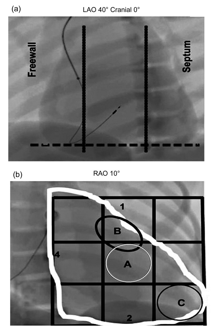
Fig. 1.
Radiological anatomy of mid-septum access
(a) 40° left anterior oblique (LAO) fluoroscopy with the lead in septal position. The tip of the lead is geared for the column, in a direction opposite to the freewall of the right ventricle (RV). (b) Septal position is represented in the 10° right anterior oblique (RAO) fluoroscopy. The letter A (white circle) represents the middle portion of the interventricular septum. The letter B (black circle) represents the high septal region and the letter C (black circle) the tip of RV. The figures are reproduced with permission from Lieberman et al. (2004)

