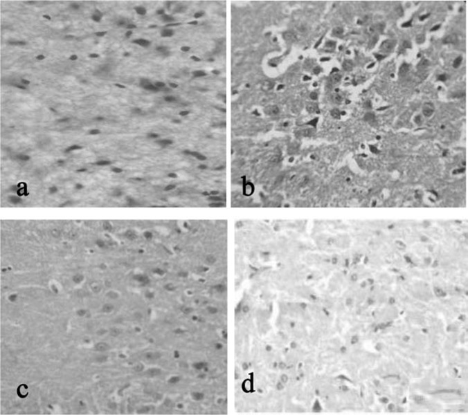
Fig. 1:

Histopathological tissue section from mid brain of rat stained with H & E in 40X. a (CONTROL): Normal architecture of brain observed. b (STRESS INDUCED): rare normal cells present in edematous cortical region with scattered damaged neurons.c(STRESS+DRUG TREATED): more normal cells are present in edematous appearing cortical region when compared to that of group II. d(drug treated): drug control animal showing normal architecture of cell as that of control.
