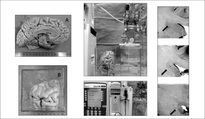
Fig. 4:
Ex-vivo infusion specimen and apparatus. 4A) External anatomy of the left hemisphere formalin fixed specimen. 4B) Specimen from 4A tailored to allow simulation of a transfrontal approach to the target. Note the olfactory bulb inferior to the letter at the picture’s top right. 4C) Priming of the neurocatheter system in fluid and measuring for planned targeting for depth of infusion. Catheter was inserted with an initial flow rate of 1.0 microliter per minute documented by indicator dye in the right-sided setup tank. 4D) Infusion catheter system at the end of the prescribed 100 microliter infusion indicating a line pressure over 500 mmHg and “infusion complete” status. 4E) Pathological sectioning showing the catheter trajectory and termination in grey matter without any evidence of indicator dye present.

