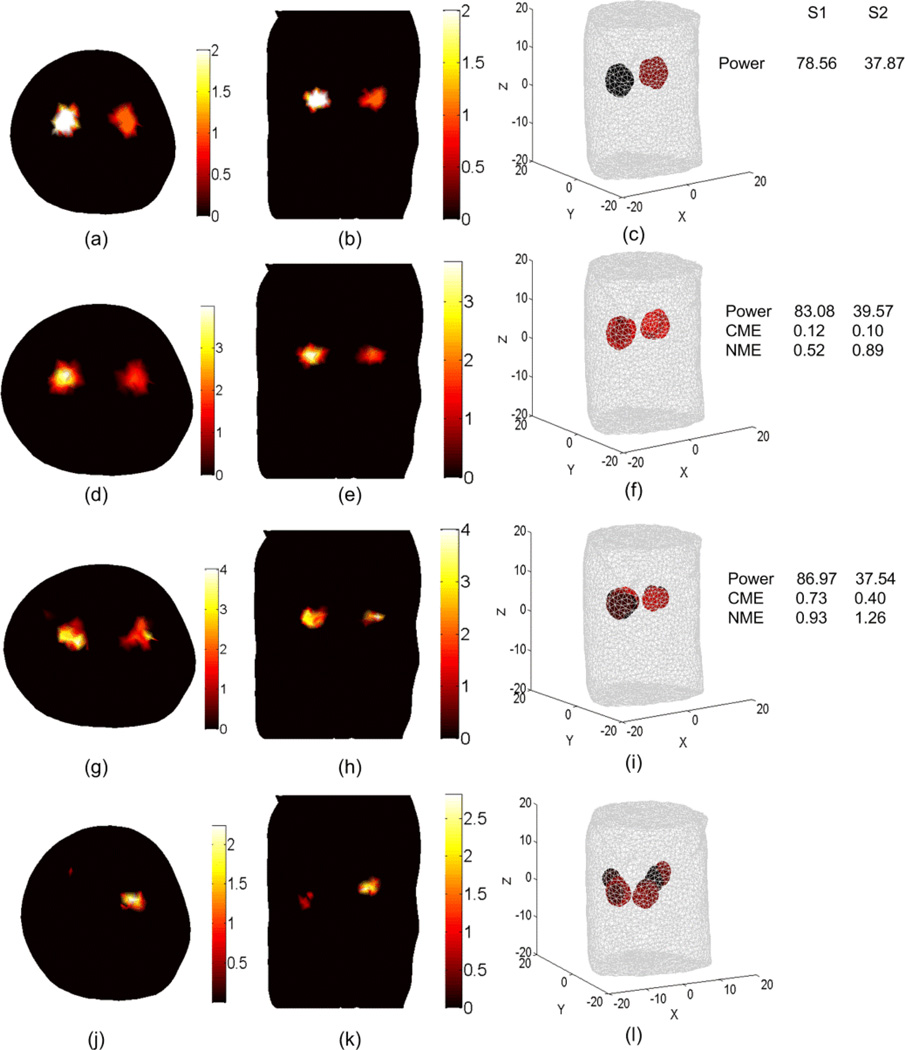
Figure 10.
BLT reconstruction of two uniform bioluminescence sources localized in the left and right kidneys (4 mm diameters). The “anatomy” is shown in Figure 3. The actual sources in transverse section (a), coronal section (b), and 3D view (c) have magnitudes 2 nW/ mm3 and 1 nW/ mm3 for the left and right source respectively. The source-detectors setup used for the DOT and BLT is as shown in Figure 3(c) where one view of a 260 degree field of view is used. The reconstructed sources are shown in (d), (e), and (f) using the DOT results shown in Figure 7 (a), (b), and (c) where segmentation using CT-scan is used. The BLT results shown in (g), (h), and (i) are obtained using unsegmented DOT results shown in Figs 4 and 5 third row. The BLT results shown in (j), (k), and (l) are obtained using DOT results assuming homogenous optical properties where there is only one scattering and absorption coefficients used for all tissues.

