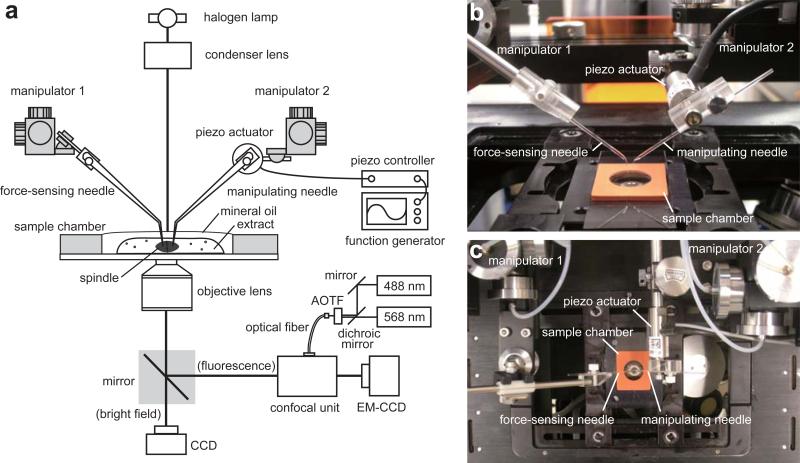
Figure 1.
Experimental set-up for analysis of metaphase spindle micromechanics. (a) Schematic of the system. A pair of microneedles is held by micromanipulators mounted on an inverted microscope. The CSF-arrested extract containing metaphase spindles is placed in a sample chamber. Confocal fluorescence imaging is used for visualizing chromosomes and spindle microtubules. Brigit field imaging is used for tracking motion of the microneedle tips. Photographs of a side view (b) and a top view (c) of the set-up around the sample chamber are presented.

