Fig. 3.
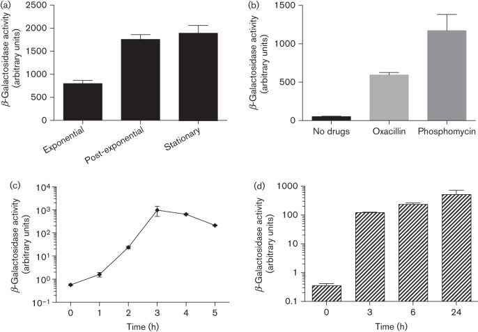
Analysis of ctpA expression. (a) The USA300 ctpA–lacZ reporter fusion strain was grown in TSB and samples were taken for β-galactosidase assays at 3 h (exponential phase), 6 h (post-exponential phase) and 10 h (stationary phase). (b) Overnight cultures of USA300 ctpA–lacZ containing subinhibitory concentrations of antibiotics were used for β-galactosidase assays. (c) The USA300 ctpA–lacZ reporter fusion strain was grown in TSB for 3 h and used to inoculate human serum. Samples were taken for β-galactosidase assays at various time points post-inoculation. The 0 h time point corresponds to 3 h growth in TSB. (d) RAW264.7 cells were infected with the USA300 ctpA–lacZ reporter fusion strain and samples were taken for β-galactosidase assays at various time points post-infection. All data shown are the mean of three independent replicates, with error bars representing ±sd.

