Fig. 5.
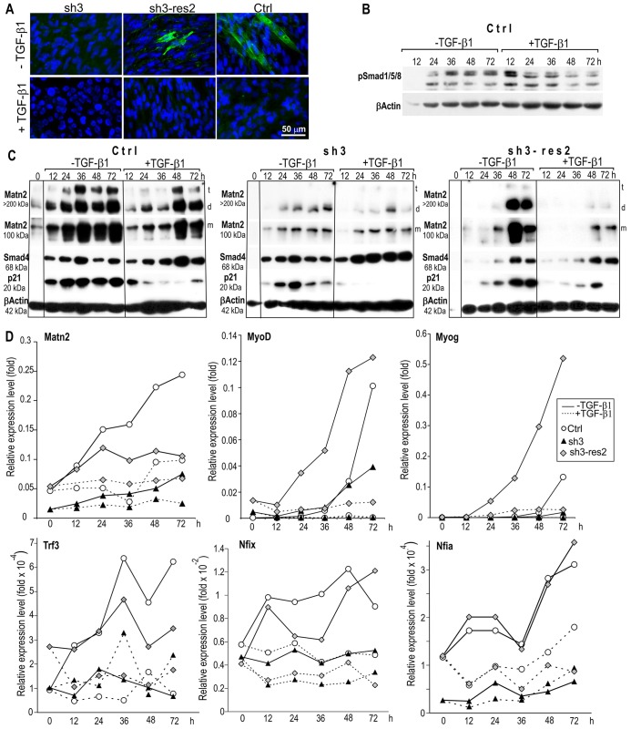
The effect of TGF-β1 on the differentiation of Ctrl, sh3 and sh3-res2 myoblasts. Cells were cultured in differentiation medium with and without TGF-β1. (A) Immunofluorescent staining for α-actinin on day 3 of differentiation. (B) Smad1/5/8 phosphorylation during the differentiation of the control cell line was assessed by immunoblotting. (C) Western blot analysis of three pooled samples for Matn2, Smad4 and p21 expression. Cytoskeletal β-actin served as a loading control. t, trimer; d, dimer; m, monomer. (D) QRT-PCR of three pooled parallel cultures using the SYBR green protocol.

