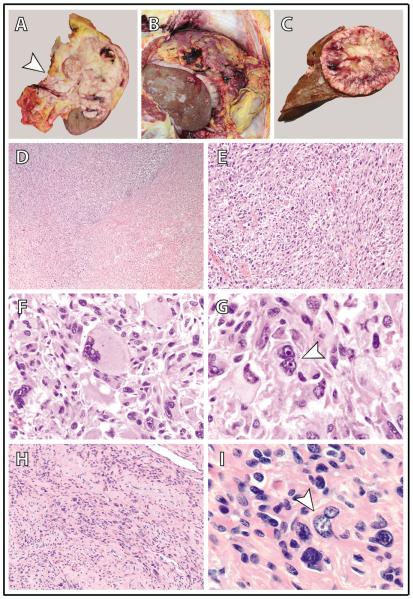
Figure 2.
Gross autopsy and histomorphologic findings in a patient with hereditary leiomyomatosis and renal cell carcinoma (HLRCC) and a high-grade, metastatic renal cell carcinoma (RCC). (A) The upper pole and middle of the left kidney were diffusely involved by a tan-white, infiltrative tumor that extended into the renal sinus and through the renal capsule into the perinephric adipose (arrowhead = renal sinus). (B) Diffuse intraperitoneal tumor metastasis (peritoneal carcinomatosis) with omental tumor caking. (C) A solitary large tumor metastasis in the liver. (D-G) The kidney tumor is comprised of sheets of highly malignant cells with extensive sarcomatoid (D and E; 40X and 200X magnification, respectively) and rhabdoid (F; 400X magnification) morphology; frequent admixed multinucleated tumor giants cells (arrowhead in G; 1000X magnification) are also identified. Tumor cells have enlarged nuclei with prominent eosinophilic nucleoli and peri-nucleolar clearing – the classic nuclear features of HLRCC-associated RCC. These features are readily seen in all histomorphologic patterns, including rhabdoid (F) and multinucleated tumor giant cells (G), and similar findings are present in all metastatic sites (not shown). (H,I) A grossly inapparent intramural uterine leiomyoma (100X and 1000X magnification in H and I, respectively); occasional cells have enlarged nuclei with eosinophilic nucleoli and peri-nucleolar clearing (arrowhead in I).

