Figure 3.
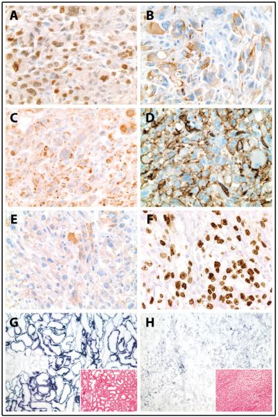
Immunohistochemistry and ancillary studies of a high-grade renal cell carcinoma (RCC) in a patient with hereditary leiomyomatosis and renal cell carcinoma (HLRCC). (A-B) Tumor cells diffusely express PAX8 (A; 400X magnification) and show patchy expression of pan-cytokeratin (B; 400X magnification). (C) Diffuse S-(2-succinyl) cysteine (2SC) staining demonstrates significant accumulation of aberrantly succinated proteins in tumor cells (400X magnification). (D, E) The hypoxia inducible factor target GLUT1 (D; 400X magnification) is strongly expressed on tumor cell membranes and shows moderate cytoplasmic staining, while CAIX (E; 400X magnification) is only weakly and focally expressed in cytoplasm of tumor cells. (F) There is diffuse nuclear accumulation of p53 in tumor cells (400X magnification). (G,H) Enzyme histochemistry for succinate dehydrogenase (SDH; 100X magnification) demonstrates significantly decreased SDH activity in tumor cells (H) compared to uninvolved contralateral right kidney (G) (intensity of staining varies directly with enzymatic activity; inset, H&E, 100X magnification).

