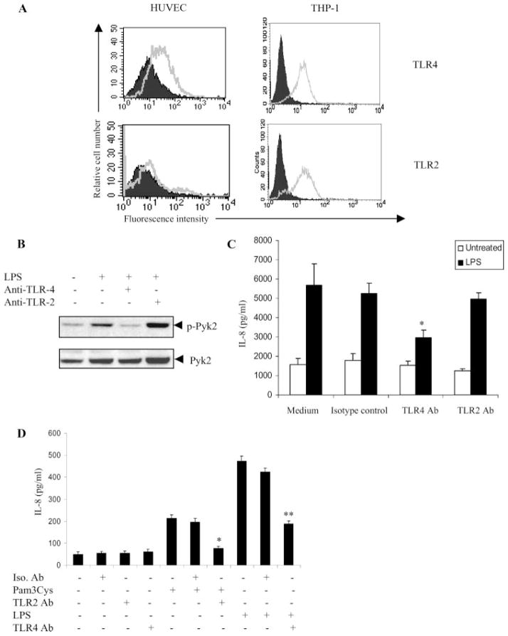
FIGURE 4.
The LPS-Pyk2-IL-8 pathway is TLR4-dependent. A, HUVEC and THP-1 were stained using Abs against TLR4 (left) or TLR2 (right) and analyzed by flow cytometry. Cells stained with control IgG represent the Ab control (AbC). Ab control (filled histogram) and TLR expression (open histogram) are indicated. B, HUVEC were pretreated with anti-TLR4 or anti-TLR2 blocking Ab (10 μg/ml) or isotype control for 1 h at 37°C. The cells were then stimulated with LPS (100 ng/ml) for 15 min in EBM with 0.5% FBS. The cells were lysed and analyzed by Western blotting with anti-phospho-Pyk2 (Tyr 402) Abs (top). The same blots were probed with anti-Pyk2 Abs (bottom). C, HUVEC preincubated with anti-TLR4 Ab, anti-TLR2 Ab, or isotype control for 1 h were cultured with or without LPS (100 ng/ml). The concentration of IL-8 in the culture supernatants was determined 24 h after stimulation. *, p < 0.05 compared with the vehicle control. Data represent the mean ± SD of three independent experiments. D, Vitamin D3-differentiated THP-1 cells preincubated with isotype control (Iso. Ab) or anti-TLR2 Ab for 1 h were cultured with or without Pam3Cys (10 pg/ml). Similarly, THP-1 cells, preincubated with isotype control (Iso. Ab) or anti-TLR4 Ab for 1 h were cultured with or without LPS (100 ng/ml). The concentration of IL-8 in the culture supernatants was determined 24 h after stimulation. *, p < 0.05 compared with the isotype control. **, p < 0.05 compared with the isotype control.

