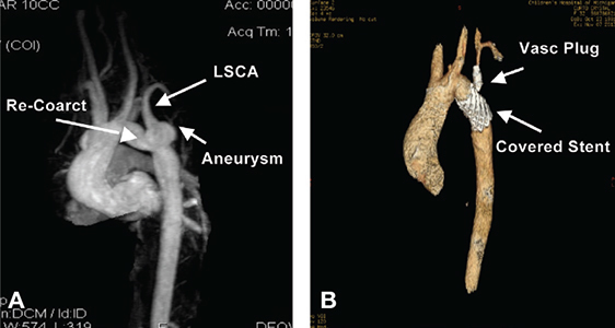
Figure 6.
(A & B) 3-dimensional reconstructive images of pre- and postcovered stent treatment of both recoarctation and aneurysm at the base of the LSCA. Figure B shows successful elimination of both the coarctation segment and aneurysm with a Cheatham-Platinum covered stent. Placement of a vascular plug was performed in the proximal LSCA following distal left common carotid to distal LSCA jump graft placement performed the previous day. LSCA: left subclavian artery.

