
Figure 4. Building a tertiary interaction.
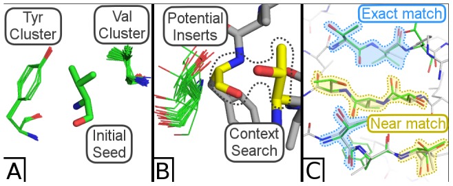
(A) Three strands are seeded by searching on a valine, which reveals two nearby clusters of valine and tyrosine. (B) Strands are extended one residue in each direction by searching for pairs of residues (colored yellow) in the context of an insertion site, yielding clusters of potential inserts (colored green). (C) The final backbone fragment (green) is fed to MadCaT, which identifies multiple compatible scaffolds. One such scaffold (PDB ID = 1E54, colored light grey) possesses many exact residue/rotamer matches to the assembled fragment (blue highlights) and many close matches (yellow highlights) that differ by a related residue (threonine to serine or valine to isoleucine) or by varying the rotamer.