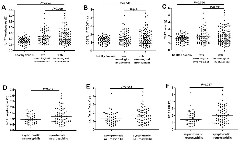
Figure 1. Increased IL-17+ and Th17 cells in peripheral bloods of neurosyphilis patients.
Peripheral blood mononuclear cells (PBMC) were stimulated and stained for flow cytometric analysis. Lymphocytes were gated according to forward and side scatter characteristics, and CD4+ T cells were gated based on CD3 and CD4 expression. (A) The percentage of total IL-17+ cells in gated lymphocytes in peripheral bloods of healthy donors (n = 70), syphilis patients without neurological disorders (including primary, secondary, latent and serofast syphilis; n = 69), and syphilis patients with neurosyphilis (n = 103) (including both asymptomatic and symptomatic neurosyphilis). (B) The percentage of Th17 cells in gated CD3+ T cells in peripheral bloods of the three groups shown in (A). (C) The percentage of Th17 cells in gated CD4+ T cells in peripheral bloods of the three groups shown in (A). (D) The percentage of total IL-17+ lymphocytes in gated lymphocytes in peripheral bloods of asymptomatic (n = 40) and symptomatic (including meningovascular, paretic, ocular and tabetic, n = 63) neurosyphilis patients. (E) The percentage of Th17 cells in gated CD3+ T cells in peripheral bloods of the two groups as in (D). (F) The percentage of Th17 cells in gated CD4+ T cells in peripheral bloods of the two groups as in (D). Each dots represents one individual. Results represent the median + individual values.

