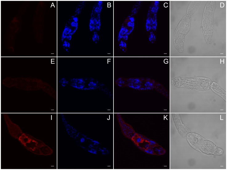
Figure 6. SmHsf1 may be localized to the acetabular glands in S. mansoni cercariae.
(A–L) Single, representative confocal sections of cercariae. A custom, rabbit polyclonal primary antibody against S. mansoni Heat shock factor 1 protein (SmHsf1) and a donkey anti-rabbit Alexa 647 secondary antibody were used to detect SmHsf1 in cercariae. (A–D) No primary negative control. The anterior region (mouth) is located near the bottom of the panels. (E–H) Pre-immune serum IgG negative control. The anterior region is located to the left. (I–L) Anti-SmHsf1. In panel I, SmHsf1 is localized to the acetabular glands (red) which traverse the entire head of the cercariae from the posterior (left) to anterior (bottom right). Panels B, F, and J are stained with DAPI. Panels C, G, and K are merged Alexa 647 and DAPI images. Panels D, H, and L are Differential Interference Contrast (DIC) images for each treatment. Scale bar, 10 µm.

