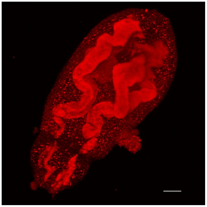
Figure 7. Antibody raised against SmHsf1 localizes to the acetabular glands extending the entire length of the S. mansoni cercarial head.

Immune staining, as in Figure 6, was used to localize anti-SmHsf1 signal (red) to acetabular glands of the S. mansoni cercariae. The anterior (mouth) is to the bottom left of the image. The image is a maximum confocal projection, and the magnification is with a 63×/1.2 W objective. Scale bar, 10 µm.
