Fig. 1.
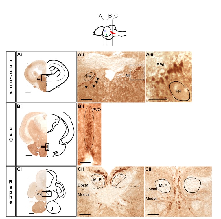
Populations of serotonin-immunoreactive (5-HT-ir) cells in the Astatotilapia burtoni male brain. The illustration at the top shows a lateral view of the brain with approximate locations of coronal sections (A–C) and corresponding 5-HT-ir populations in dorsal and ventral periventricular pretectal nuclei (PPd/v, blue), nucleus of the paraventricular organ (PVO, green) and raphe (red). Representative low magnification photomicrographs of coronal sections in the PPd/v (Ai), PVO (Bi) and raphe (Ci) are shown on the left and outlined as a mirror image on the right; labeled boxed areas are shown at higher magnification in Aii, Bii and Cii. In Aii, 5-HT-ir cells (arrowheads) in the PPv line the ventral side of the fasciculus retroflexus (FR) fiber bundle and 5-HT-ir cells in the PPd (boxed area) are shown at higher magnification in Aiii. We used the base of the medial longitudinal fasciculi (MLF) fiber tracts as landmarks to trace the boundary (black dashed line) between dorsal and medial raphe subregions, depicted in Cii and Ciii, raphe coronal sections that were 120 μm apart. In the more anterior section (Cii), dorsal raphe 5-HT-ir cells can be seen densely packed surrounding the MLF fiber tracts and medial 5-HT-ir cells are more scattered. The more posterior section (Ciii) shows 5-HT-ir cells arranged in parallel, on both sides of the midline. Scale bars: Ai–Ci, 500 μm; Aii, Bii, Cii and Ciii, 100 μm; Aiii, 50 μm.

