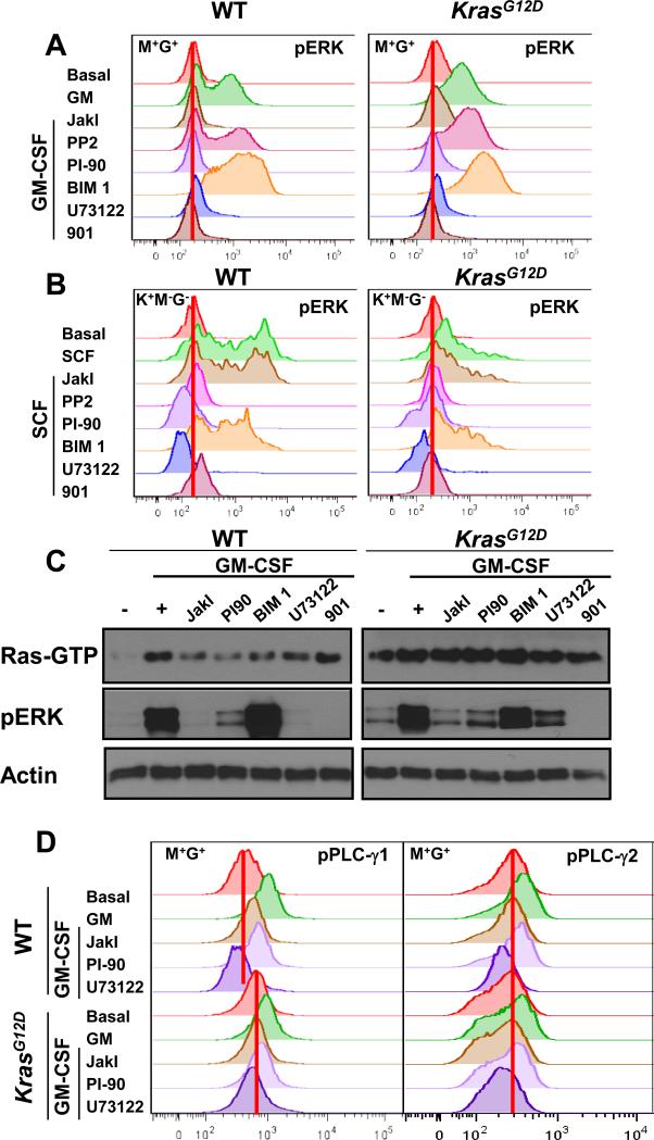
Fig. 1. PI-90 and U73122 impair Ras and ERK activation in wild-type and Mx1-Cre, KrasG12D hematopoietic cells and reduce PLC-γ phosphorylation.
(A) M+G+ cells from wild-type (WT) and Mx1-Cre, KrasG12D (KrasG12D) mice were incubated for 30 min with the indicated inhibitors (each at final concentration of 5 μM), stimulated with GM-CSF for 10 min, and then analyzed by phospho-flow cytometry to determine the abundance of pERK. Basal indicates cells that were treated neither with inhibitor nor cytokine. The red vertical lines indicate basal median fluorescence intensity (MFI) values. (B) K+M−G− cells from WT and KrasG12D mice were incubated for 30 min with the same inhibitors used in (A) before they were stimulated with SCF for 15 min and then analyzed by phospho-flow cytometry to determine pERK abundance. (C) Ras-GTP and pERK abundances were measured in bone marrow cells from WT and KrasG12D mice (as described in the Methods) that were treated with the indicated inhibitors before being stimulated with GM-CSF. Actin was used as a loading control. (D) The amounts of phosphorylated PLC-γ1 (pPLC-γ1) and PLC-γ2 (pPLC-γ2) were determined by flow cytometric analysis of WT and KrasG12D mouse bone marrow M+G+ cells that were pretreated with the indicated inhibitors before being stimulated with GM-CSF. Data from additional independent experiments are shown in figs. S2 B and C, and S3.

