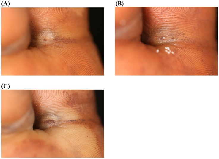
Figure 3. Photo series of a lesion located at the base of the first toe; treatment with KMnO4.
(A) Baseline: A lesion in stage IIIa with a diameter of 10 mm at the base of the first toe. The abdominal cone is the circular brownish protrusion in the center of the elevation. The dermal papillae next to the lesion contain faecal material expelled by the parasite. (B) Day 3: The sand flea has expulsed several eggs (white oval dots). One of the eggs is in progress of being expelled. The appearance of the lesion has not changed. (C) Day 7: The lesion has retained its size and remains elevated. Recently excreted faecal material has spread into the dermal papillae next to the lesion, another indicator that the parasite remained viable.

