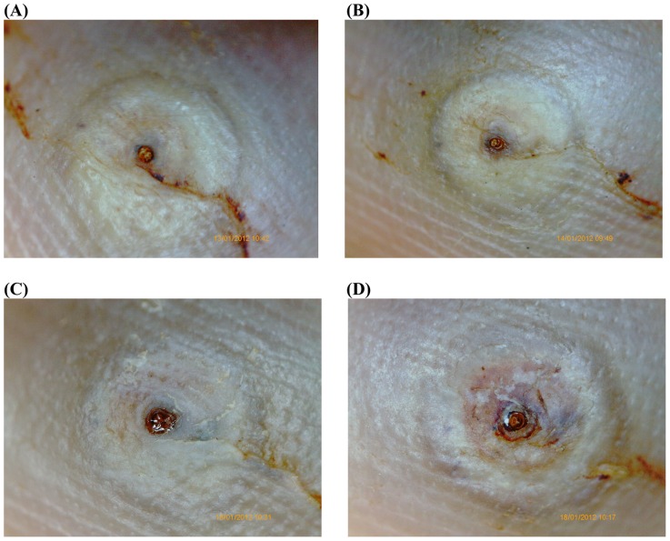
Figure 5. Photo series of a lesion documented by the digital handhold video microscope at 200 fold magnification; treatment with KMnO4.
(A) Baseline: Lesion in stage IIIa. The abdominal cone is the circular brownish protrusion surrounded by the characteristic watchglass-like elevation. The curved line is faecal material of the parasite that has spread into dermal papillae. (B) Day 3: The embedded parasite has grown slightly and the convex elevation is more embossed. The abdominal cone is still brownish and shining. (C) Day 5: The appearance of the lesion has not changed. Faecal liquid is excreted through the abdominal cone and appears as a clear, light-reflecting “pond” on the top of the cone. (D) Day 7: The abdominal cone is still brownish and shining. The lesion has a convex double-rim appearance. Two viability signs (pulsation of the parasite and excretion of liquid) were present at this moment.

