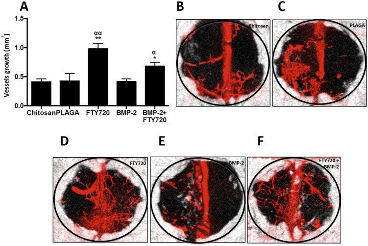
Figure 7. FTY720 enhances vascularization in a critical size cranial defect.
(A) Vascularization in the critical size cranial defect measured using microfil enhanced microCT imaging. (B–F) Volume in the defect region occupied by vessel ingrowth 9 weeks after different treatments (** p<0.01 & *p<0.05 compared to chitosan control; αα p<0.01 & α p<0.05 compared to PLAGA control) MicroCT images showing vessel and bone in-growth at week 9 for animals treated with (B) Chitosan, (C) Chitosan + PLAGA microspheres, (D) Chitosan + PLAGA microspheres loaded with FTY720, (E) Chitosan loaded with BMP-2 and (F) Chitosan loaded with BMP-2 + PLAGA microspheres loaded with FTY720.

