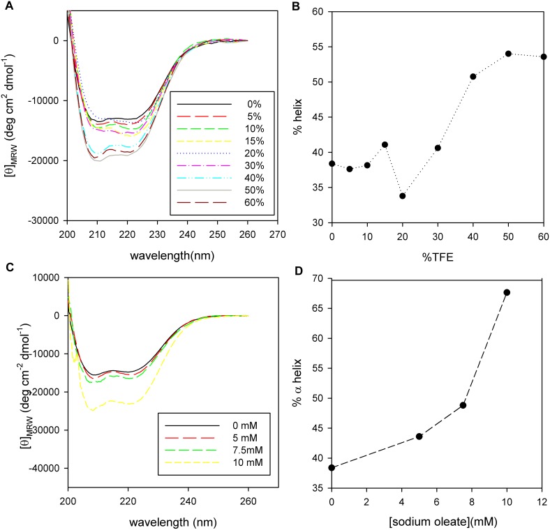
Figure 4. PhaPAz secondary structure in different environments.
CD Spectra of PhaPAz (3 µM) in sodium phosphate buffer (20 mM, NaCl 50 mM, pH 7.3, 1 mM DTT) (A) at different TFE concentration (from 0 to 60%) or (C) at different sodium oleate concentrations (0 to 10 mM). α-helix content, calculated using K2D3 algorithm, as a function of TFE (B) or sodium oleate (D).

