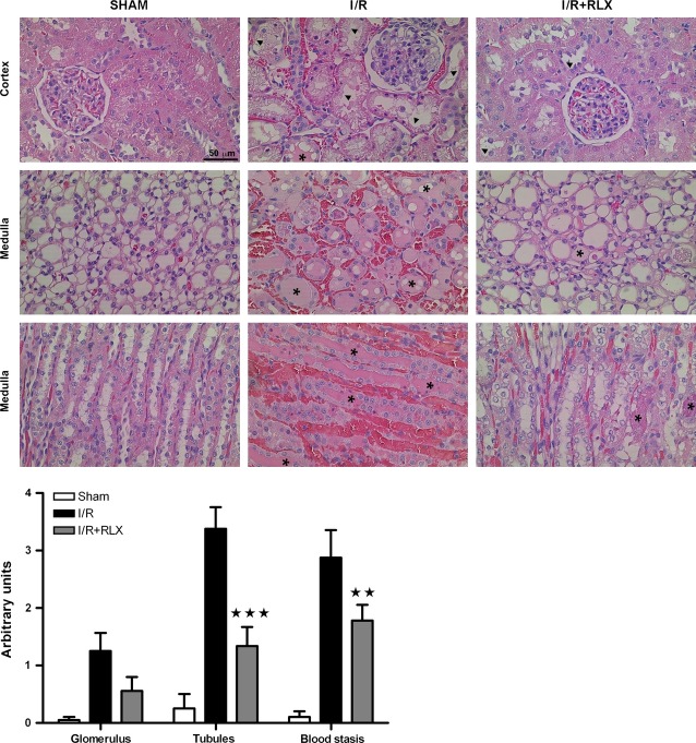
Figure 2.
Representative histopathological features of kidney biopsies in the different experimental groups and semi-quantitative assessment of the severity of kidney damage. Upper panels: widespread tubular cell vacuolization, shedding of the tubular epithelial lining (arrowheads) and hyaline tubular casts (asterisks) are seen in the renal cortex and medulla; the interstitial connective tissue shows dilated microvessels filled with blood and sparse haemorrhage foci. Below panel: severity scoring of the histological damage. Significance of differences: ⋆⋆P < 0.01 and ⋆⋆⋆P < 0.001 versusIR.

