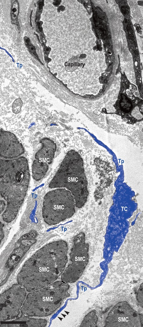
Figure 3.

Muscular layer of human oesophagus. Transmission electron microscopy. Telocyte (TC) with two visible Telopodes (Tps) in close vicinity of smooth muscle cells (SMC), and wrapping them. Fragments of (most probably) the same Tp are interposed between SMC and a blood capillary. Small electron-dense nanostructures (arrow-heads) are seen between both membranes – SMC and Tp.
