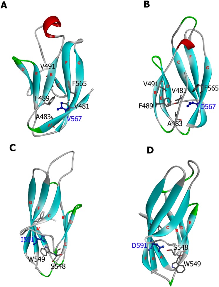
Figure 8. Structural comparison of the predicted wild-type and mutant α-DG Ig-like domains.
The wild-type zebrafish (Panel A), the zebrafish V567 mutant (Panel B), the wild-type murine (Panel C) and the murine I591D mutant (Panel D) models are shown using their corresponding average structure of the last 25 ns simulation. The location of the residues described in the current study and strands A′, B, C, D, E, F, and G are also labeled.

