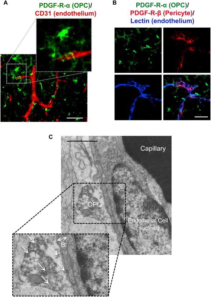
Figure 3. OPC-endothelium interaction.
A. Immunostaining showed that some OPCs (green: PDGF-R-α) are located closely to cerebral endothelium (red: CD31) in mice at post-natal day 0–1. Scale bar = 50 µm. B. Triple staining showed that there is little overlap between PDGF-R-α and PDGF-R-β. Scale bar = 10 µm. C. Electron micrography also confirmed that OPCs attached directly to the basal lamina (Bl) of endothelial cells in corpus callosum. arrows: PDGF-R-α positive signals, Scale bar = 1 µm.

