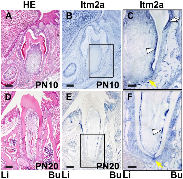
Figure 4. The in situ expression of Itm2a mRNA in the developing tooth germ at the tooth root formation and the tooth eruption stages.
A. An HE-stained section shows the tooth germ on PN10 at the tooth root formation stage. B. The in situ signal of Itm2a was observed in the tooth germ on PN10. C. The boxed area in B is shown at a higher magnification. The in situ expression of Itm2a was detected in the ameloblasts (white arrow), odontoblasts (white arrowhead) and HERS (yellow arrow). D. An HE-stained section shows the tooth germ on PN20 at the tooth eruption stage. On PN20, the root formation is almost completed, and the crown is exposed. E. The Itm2a expression was observed in the odontoblasts and HERS. F. The boxed area in E is shown at a higher magnification. The in situ expression of Itm2a signal was detected in the odontoblasts (white arrowhead) and HERS around the root apex (yellow arrow). Li; lingual side, Bu; buccal side. Scale bars; 200 µm (A, B, D, E), 100 µm (C, F).

