
Figure 6.
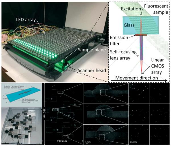
Flatbed scanner converted to a 2.2 giga-pixel fluorescent imaging device with a field of view of 532 cm2. (Top left) Photograph of the device consisting of a flatbed scanner fitted with an emission filter and an array of 600 LEDs providing uniform illumination over the entire field of view. (Top right) Diagram showing the light path inside the scanner head. The excitation arrives over a 45 degree angle to minimize the amount of light entering the scanner's gradient index lens. The light scattered by the sample is attenuated by the filter, while the emitted fluorescent light passes through it. (Bottom) Experiment designed to validate the performance of the system. Microfluidic chips filled with whole blood spiked with 10μm fluorescent particles are scattered over the field of view. Scanned image of the sample and (from left to right) increasing zooms to focus onto the chambers. Last column shows fluorescent microscope comparison images captured by a 4x objective-lens (NA=0.13). This scanner-based fluorescent imaging device achieved 98.8% accuracy with ~1% standard deviation for counting the particles inside the chambers. The whole field of view, 532 cm2, can be imaged in <5 minutes. Reproduced from Ref. 32 with permission from The Royal Society of Chemistry.