
Figure 2.
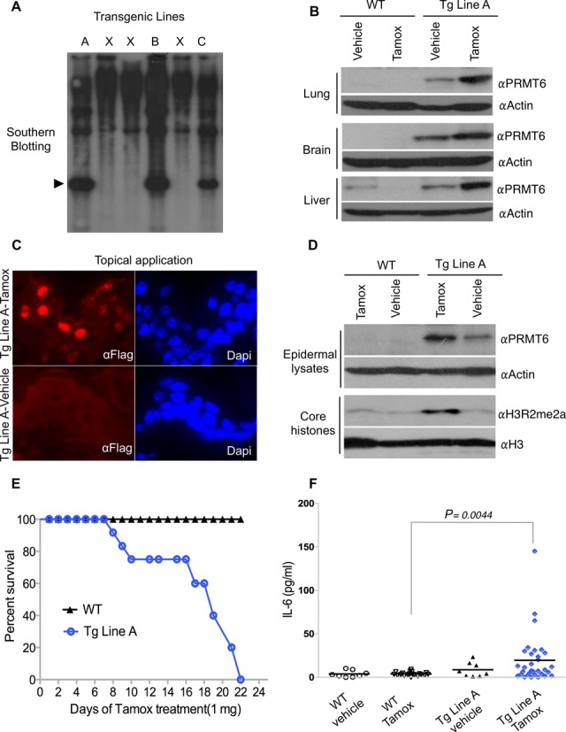
The inducible ER*-PRMT6 fusion is functional in vivo. (A) Three transgenic lines were established using the pCAGGS-ER*-PRMT6 vector. Southern analysis was performed with a PRMT6 cDNA probe. The arrowhead indicates the transgene. (B) Intraperitoneal injection of 1-mg Tamox, for 5 consecutive days, induced stabilization of ER*-PRMT6 in lung, brain and liver of Line A mice. The vehicle (sunflower oil) was injected as a control. (C) IF for the Flag-tag performed on skin sections from transgenic Line A mice shows ER*-PRMT6 stabilization in the nucleus when Tamox was applied topically on shaved dorsal skin (1 mg/day) for 5 days. (D) Core histones were isolated from epidermal scrapes of topically Tamox-treated mice (1 mg every other day for 10 weeks) and subjected to WB with an αH3R2me2a antibody. (E) The ER*-PRMT6 Line A mice die upon prolonged Tamox administration. A survival curve was generated after topical Tamox administration (1 mg/day) until death of all transgenic mice occurred (n = 14). WT mice were not impacted by this treatment. (F) IL-6 serum levels after 12 topical applications of Tamox (1 mg/day) were measured by ELISA in four groups of animals (WT, WT Tamox-treated, Transgenic (Tg) Line A, Tg Line A Tamox-treated). Wilcox and Student t-tests were run and a P value of 0.004 was obtained for the Tg Tamox-treated group (n = 32) when compared with the WT Tamox-treated group (n = 20) for both tests. All the mice in this study were healthy at the time of serum harvesting. All mice were between 8 and 9 weeks of age, and females and males were equally represented.