
Figure 5.
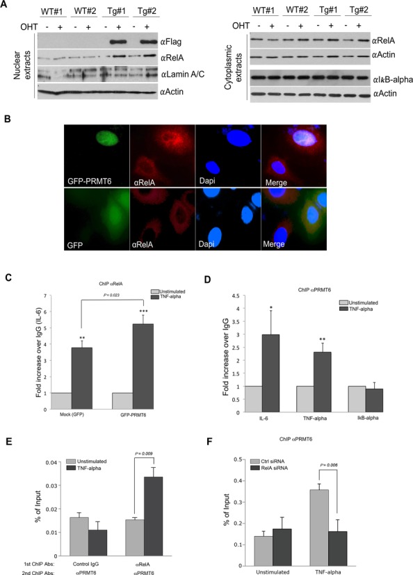
PRMT6 promotes RelA shuttling into the nucleus and ChIPs at a subset of NF-κB-regulated promoters. (A) Primary MEFs were isolated from WT and ER*-PRMT6 mice, cultured for 3 days and treated with OHT for 2 weeks. Cell fractionation was performed. Western analysis for the Flag-tag shows stabilization of ER-PRMT6 in the nuclear fraction, corresponding to increased nuclear RelA. WB for RelA and IκB-α on the cytoplasmic extracts shows no change in the levels of these two proteins. (B) Immunofluorescence using a αRelA antibody following transient transfection of GFP-PRMT6 in HeLa cells shows increased nuclear RelA in transfected cells. A GFP-expressing construct was used as negative control. (C) HeLa cells were transiently transfected with a GFP or GFP-PRMT6 expressing vector for 24 h, at which time cells were left untreated or treated with TNF-α (10 ng/ml, 30 min). ChIP analyses at the κB consensus region of IL-6 promoter were then performed using αRelA antibody. Values were normalized to an intergenic region 8000 bp upstream IL-6 consensus sequence. For each transfection, the mean value of the unstimulated group was arbitrarily set as 1. Error bars represent standard deviations calculated from triplicates. The P values of the TNF-α-stimulated groups compared to the unstimulated are 0.0024 and 0.0003 for the Mock and GFP-PRMT6-transfected cells, respectively. The P value of the fold increase difference for PRMT6-overexpressing cells versus the Mock is 0.023. (D) HeLa cells were grown to 90% confluency, at which point cells were left untreated or treated with TNF-α (10 ng/ml, 30 min). ChIP analyses at the κB consensus region of IL-6, TNF-α and IκB-α promoters were then performed using αPRMT6 antibody. Values were normalized to an intergenic region 8000 bp upstream IL-6 consensus sequence. Error bars represent standard deviations calculated from triplicates. The mean value of the unstimulated groups was arbitrarily set as 1. The P values of the TNF-α-stimulated groups compared to the unstimulated are 0.024, 0.0053 and 0.84 for IL-6, TNF-α and IκB-α genes, respectively. (E) The recruitment of PRMT6 and RelA to the IL-6 promoter consensus region was evaluated by ChIP/re-ChIP experiment. HeLa cells were either left unstimulated or treated with TNF-α (10 ng/ml, 30 min) before the ChIP/re-ChIP experiments were performed using indicated antibodies. The ChIP DNA was analyzed by qPCR with primers for the IL-6 consensus region. (F) ChIP experiment was performed using αPRMT6 antibody in control and RelA-knockdown HeLa cells, which were either left unstimulated or treated with TNF-α.