
Figure 4.
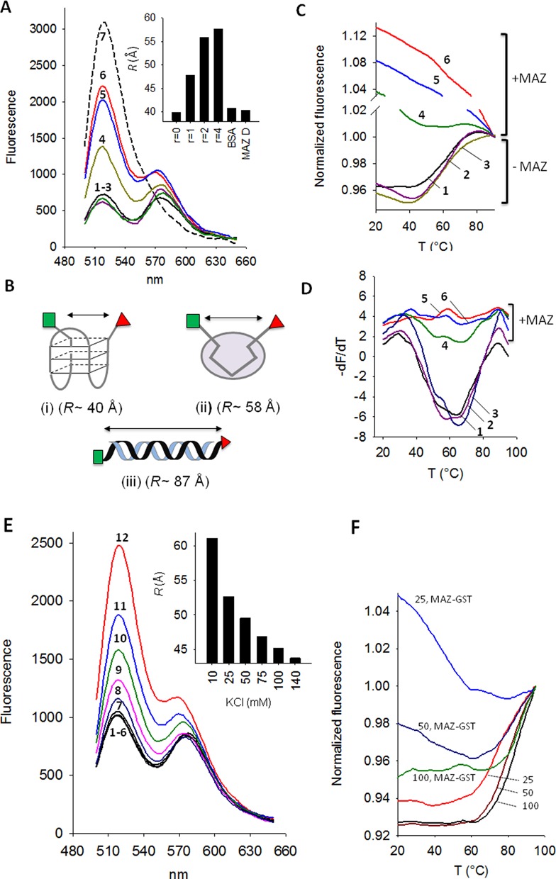
(A) Fluorescence emission of 200 nM quadruplex hras-1 tagged at the 5′ and 3′ ends with FAM and TAMRA in 50 mM Tris-HCl, pH 7.4, 50 mM KCl, Ex = 480 nm. Spectra: 1, F-hras1-T; 2, F-hras1-T + heated MAZ-GST (1:4); 3, F-hras1-T + BSA (1:4); 4–6, F-hras1-T + MAZ-GST at r = 1, 2 and 4, respectively; 7, F-hras1-T hybridized to complementary 27Y strand to give the duplex. Each quadruplex–protein mixture has been incubated for 2 h before FRET analysis. The inset shows the end-to-end distance (R, Å) of quadruplex hras-1 treated for 2 h with MAZ-GST (r = 0, 1, 2 and 4), with BSA (r = 4) and with denatured MAZ-GST (r = 4) (MAZ D); (B) cartoon showing the three forms of hras-1 and for each the calculated end-to-end distance; (C) Normalized fluorescence versus T curves of quadruplex hras-1 in 50 mM Tris-HCl pH 7.4, 50 mM KCl: curve 1, quadruplex hras-1; curve 2, quadruplex hras-1 treated for 2 h with heated MAZ-GST (r = 4); curve 3, quadruplex hras-1 treated for 2 h with BSA (r = 4); curves 4, 5 and 6, quadruplex hras-1 treated for 2 h with MAZ-GST at r = 1, 2 and 4, respectively; (D) –dF/dT versus T curves as in C; (E) FRET of hras-2 tagged with FAM and TAMRA (F-hras2-T) in 10, 25, 50, 75, 100, 140 mM KCl (curves 1–6) and after treatment with MAZ-GST (r = 4) (curves 12 to 7). The inset shows the decrease of the end-to-end distance of quadruplex hras-2 treated with MAZ-GST (r = 4) at various KCl concentrations; (F) Normalized fluorescence versus T curves of quadruplex hras-2 in 25, 50 and 100 mM KCl with and without treatment with MAZ-GST (r = 4) for 2 h.