
Figure 4.
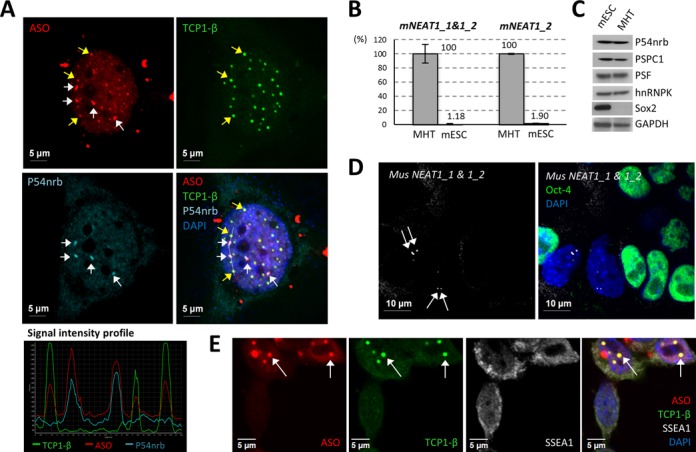
PS bodies and paraspeckles are distinct ASO-containing subnuclear bodies. (A) Transfected ASOs can co-localize with either PS body protein TCP1-β or with paraspeckle protein P54nrb in HeLa cells. White or yellow arrows indicate the co-localization of PS-ASOs with P54nrb in paraspeckles or TCP1-β in PS bodies, respectively. The signal intensity profile of several ASO-containing structures (marked with a dash line in the merge panel) confirmed the co-localization of the ASOs with either TCP1-β or P54nrb. (B) NEAT1 RNA was barely detectable by qRT-PCR in mESCs, as compared with the expression level in control MHT cells. Relative levels of NEAT1_1&1_2 and NEAT1_2 in MHT cells and mESCs are indicated. The error bars represent standard deviation from three parallel experiments. (C) Major paraspeckle proteins were expressed comparably between mESCs and MHT cells. Sox2 served as a mESC marker. GAPDH served as a loading control. (D) RNA-FISH of NEAT1 RNA showed no paraspeckle formation in mESCs. Oct-4 protein staining served as a mESC marker. Arrows indicate paraspeckles in differentiated cells. (E) PS bodies can form in mESCs lacking paraspeckles. Arrows indicate the co-localization of TCP1-β and ASOs in PS bodies. SSEA1 served as a mESC marker.