
Fig. 7A–B.
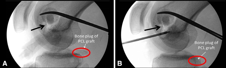
Fluoroscopic images of the graft passage are shown. (A) The tibial inlay bone plug I shown before being seated. The circle surrounds the bone plug and the black arrow points to the femoral tunnels. (B) The PCL graft is seated into the tibial socket. The circle surrounds the same bone plug now reduced into the tibial socket and the black arrow points to the femoral tunnels. (Adapted from Weber and Sekiya [55], with permission from Saunders, Elsevier, Inc.)