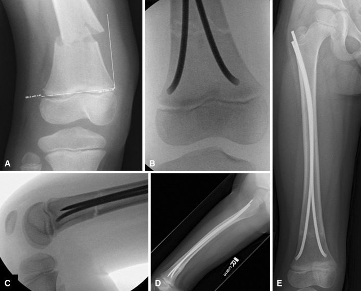
Fig. 3A–E.
(A) A supracondylar femur fracture is shown. (B, C) The fracture is treated with antegrade nails such that a spread of the nails is achieved at the fracture site and the nail tips point away from each other. (D, E) Full-length femur radiographs show the insertion of the nail below the greater trochanter with one nail C-shaped and the other S-shaped.

