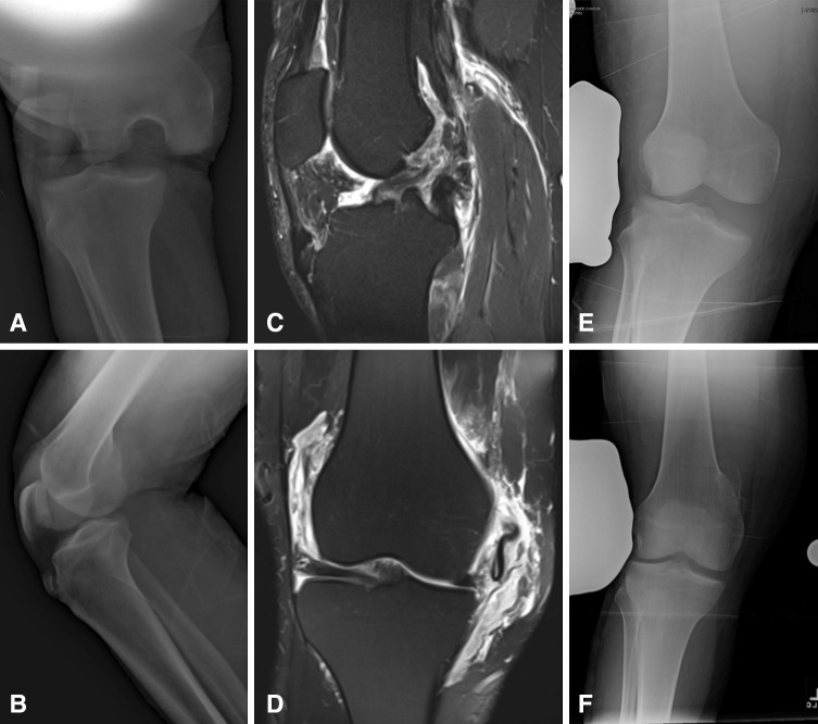
Fig. 1A–F.
Images illustrate the case of a 34-year-old dairy farmer who was kicked by a cow and sustained a KDIV knee dislocation. (A) AP and (B) lateral radiographs demonstrate the knee dislocation before reduction. (C) A sagittal short tau inversion recovery MR image and (D) a coronal fast spin echo T2-weighted MR image with fat saturation demonstrate complete ACL, PCL, lateral collateral ligament/posterolateral corner, and MCL injuries. He also had a significant posteromedial corner injury, which included medial meniscus and posterior oblique ligament tears. Valgus stress radiographs of the (E) injured and (F) uninjured knee demonstrate a greater than 10-mm side-to-side difference. (F) The uninjured knee valgus stress radiograph has been flipped horizontally to facilitate comparison with (E) the injured knee.

