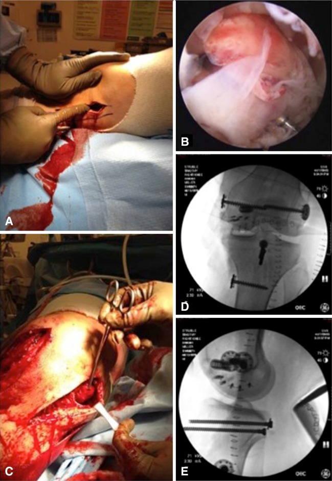
Fig. 3A–E.
Operative management of the KDIV injury in the patient depicted in Figure 1 is illustrated. (A) A medial egress incision is created. (B) Diagnostic arthroscopy confirmed a bicruciate injury. (C) Extraarticular reconstruction of the medial ligamentous injury is performed using a modified Bosworth technique. Postoperative (D) AP and (E) lateral imaging demonstrate typical hardware placement for the medial reconstruction, which is used intraoperatively to confirm an isometric location.

