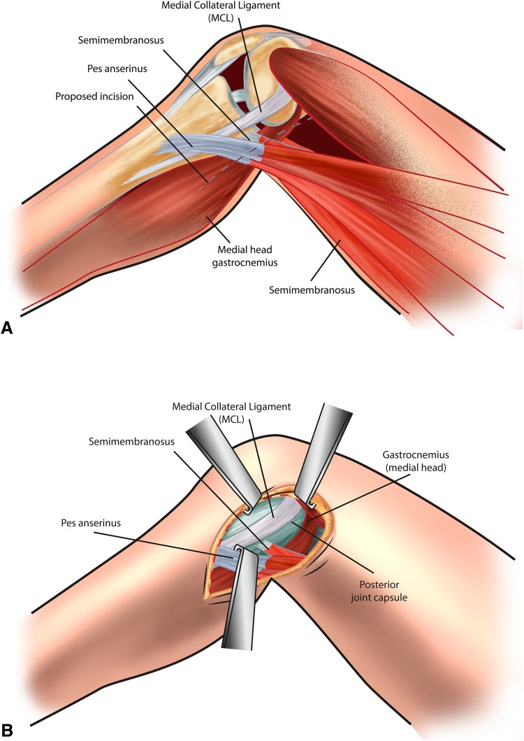
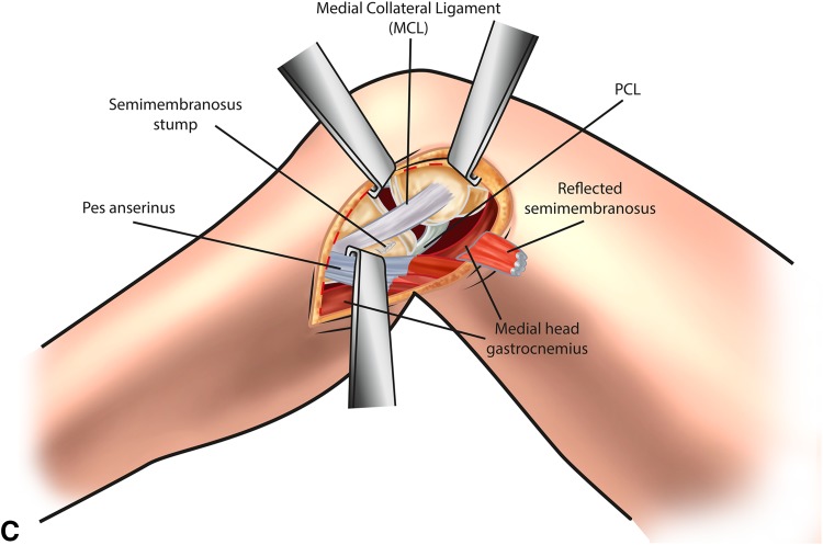
Fig. 2A–C.
(A) A skin incision is placed at the back edge of the medial tibia, coursing proximally to the posterior edge of the medial epicondyle. Superficial dissection is made through the sartorius fascia along the line of the skin incision. (B) Deep dissection is made between the posterior knee joint capsule and the gastrocnemius. Partial detachment of the semimembranosus is required to access this interval. (C) Exposure of the proximal tibia and capsulotomy allows identification of the PCL.


