
Fig. 2A–C.
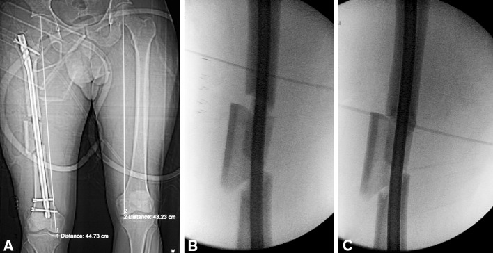
(A) A CT scan demonstrates a distraction of 1.5 cm after antergrade nailing of a Type C fracture performed on a fracture table. (B) An intraoperative fluoroscopic image shows the distracted femur fracture. (C) Using a back-slapping technique, a fluoroscopy image demonstrates that the distraction has been corrected.