Fig. 3.

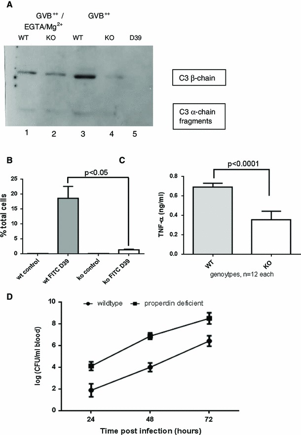
Complement activation, phagocytosis and intravascular survival of S. pneumoniae D39. a C3 reactivity of separated bacterial pellets after incubation with mouse sera (pool n = 6) under conditions favouring the alternative pathway (GVB++/EGTA/Mg2+) or classical pathway (GVB++) activation, 37 °C, 30 min (Western blot); lane 5 shows specificity of rat antimouse C3 antibody (only D39, no serum). b Pools of 9 sera-matching each cell genotype were used to opsonise FITC-labelled S. pneumoniae before addition to respective mouse splenocytes for 1 h. Flow cytometry was performed, while quenching extracellular signal with trypan blue (representative analysis of n = 2). C After 24 h’ infection of splenic macrophages with D39, there is decreased TNF-α production (measured using L929 assay) by splenocytes from properdin-deficient mice in the presence of properdin-deficient sera compared to wildtype cells with wildtype sera. d Both genotypes were injected i.v. with 10e6 D39 and bacterial counts (±SEM) determined for each of 3 days from n = 5 each genotype (p < 0.05)
