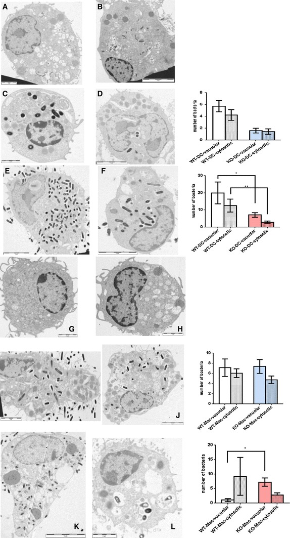
Fig. 5.

Vacuolar and cytosolic localisation of L. monocytogenes in dendritic cells (a–f) and macrophages (g–l) from wildtype and properdin-deficient mice (TEM) at 4 h (C,D,I,J) and 24 h (e, f, k, l) p.i. Typical electron micrographs are juxtaposed to quantitative analysis of macrophages and dendritic cells from both genotypes (WT-DC n = 73; KO-DC n = 12 for 4 h; WT-DC n = 11; KO-DC n = 16 for 24 h, and WT-Mac n = 26; KO-Mac n = 34 for 4 h; WT-Mac n = 5; KO-Mac n = 16 for 24 h). Note the overall increase in bacteria at 24 h. *p < 0.05; **p < 0.05 by unpaired t test; variables are significantly different (F test) for analyses of c/d, e/f, k/l
