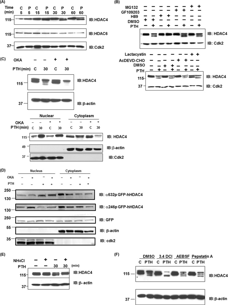
FIGURE 5.
PKA, phosphatase, and lysosomal inhibition blocks PTH-induced HDAC4 degradation. A, total cellular lysates (30 μg) isolated from 10−8 m PTH and control-treated UMR 106–01 cells for 5, 15, 30, and 60 min were used for Western blot analysis using anti-HDAC 4, 6, or Cdk2 antibodies. Anti-Cdk2 was used as the loading control. B, total cellular lysates isolated from UMR 106-01 cells, which had been preincubated with protein kinase A inhibitor H89 (50 μm) for 30 min, protein kinase C inhibitor GF109203 (5 μm) for 30 min, proteasome inhibitor MG132 (5 μm), lactacystin (5 μm), or caspase-3 inhibitor AcDEVD-CHO (100 μm) for 60 min, and then with or without PTH (10−8 m) stimulation for 30 min. Total cellular lysates were used for Western blot analysis using anti-HDAC4. Anti-Cdk2 was used as a loading control. C, total cellular lysates or nuclear/cytoplasmic extracts isolated from UMR 106-01 cells, which had been preincubated with phosphatase inhibitor, okadaic acid (OKA, 50 nm) for 60 min and then with or without PTH (10−8 m) stimulation for 30 min. Total cellular lysates or nuclear/cytoplasmic extracts were used for Western blot analysis using anti-HDAC4. Anti-β-actin was used for total cell lysates or cytoplasm as a loading control. Anti-Cdk2 was used as a nuclear loading control. D, nuclear and cytoplasmic extracts isolated from UMR 106-01 cells, which had been preincubated with phosphatase inhibitor, okadaic acid (OKA, 50 nm) for 60 min and then with or without PTH (10−8 m) stimulation for 30 min. The nuclear and cytoplasm extracts were subjected to immunoblotting with anti-phosphoSer632 hHDAC4, anti-phosphoSer246 hHDAC4, anti-GFP, anti-β-actin, and anti-Cdk antibodies. Anti-Cdk2 was used for the nucleus and anti-β-actin was used for cytoplasm as loading controls. E, total cellular lysates isolated from UMR 106-01 cells were preincubated with NH4Cl (20 mm) for 16 h and then with PTH (10−8 m) stimulation for 30 min. Total cellular lysates were used for Western blot analysis using anti-HDAC4. Anti-β-actin was used for loading control. F, UMR 106–01 cells were preincubated with vehicle, serine protease inhibitors, 3.4 DCl (50 μm) or AEBSF (200 μm), aspartic protease inhibitor pepstatin A (10 μm) for 90 min and then with or without PTH (10−8 m) stimulation for 30 min. Total cellular lysates were used for Western blot analysis using anti-HDAC4. Anti-β-actin was used as a loading control.

