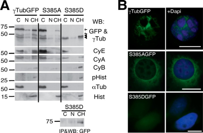
FIGURE 5.

Ser385-γ-tubGFP mutants affect the nuclear localization of γ-tubulin. A and B, U2OS cells transfected with γ-tubGFP, Ala385-γ-tubGFP (S385A), or Asp385-γ-tubGFP (S385D) were examined by Western blotting (A) and immunofluorescence microscopy (B). A, the various biochemical fractions obtained from U2OS cells were analyzed by Western blotting (WB) with anti-GFP (GFP), anti-γ-tubulin (γTub), anti-cyclin E (CyE), anti-cyclin A (CyA), anti-cyclin B (CyB), anti-phospho-histone H1 (pHist), anti-α-tubulin (αTub), and anti-histone (Hist) antibodies (n = 3) as in Fig. 2E. Anti-GFP antibody immunoprecipitates of cell lysates prepared as described in Fig. 2E but expressing Asp385-γ-tubGFP were analyzed by WB. Arrows and arrowheads indicate the GFP and endogenous γ-tubulin, respectively. B, the cellular location of γ-tubGFP, Ala385-γ-tubGFP (S385AGFP), and Asp385-γ-tubGFP (S385DGFP) was determined by immunofluorescence analysis, and nuclei were detected using DAPI (blue); scale bars, 10 μm.
