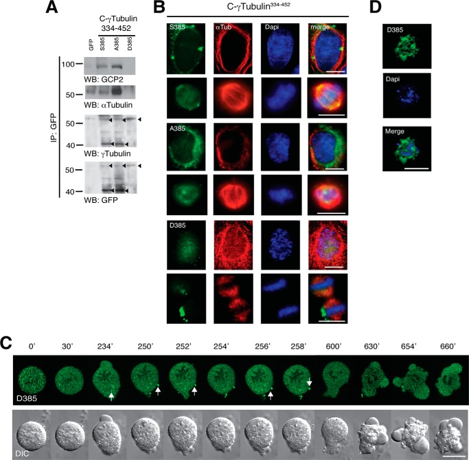
FIGURE 8.
Asp385-Cγ-tubGFP does not bind to GCP2 and α-tubulin and influence mitotic progression. A–D, U2OS cells were simultaneously synchronized in early S phase as in Fig. 1A and transfected with GFP, Ser385-Cγ-tubGFP (S385), Ala385-Cγ-tubGFP (A385), or Asp385-Cγ-tubGFP (D385). A, Western blot shows the expression of the various Cγ-tubGFP mutants. The blots were analyzed with anti-GFP antibody and sequentially stripped and reprobed with antibodies against γ-tubulin, GCP2, and α-tubulin. Arrowheads, immunoprecipitated (IP) GFP-fused proteins (n = 3). B, after the double thymidine block treatment, U2OS cells expressing the indicated constructs were released for 9 h. Localization of the Cγ-tubGFP mutants was examined by immunofluorescence staining with anti-α-tubulin (αTub; red) and nuclei DAPI staining (blue) in human U2OS cells incubated for 9 h (n = 3–5). Scale bars, 10 μm. C, differential interference contrast (DIC)/fluorescence images of time lapse from a U2OS cell with chromatin-bound Asp385-Cγ-tubGFP that arrests in metaphase. Images were collected every 2 min. The image series shows chosen frames of the location of Asp385-Cγ-tubGFP (n = 14). D, after double thymidine block treatment, U2OS cells expressing Asp385-Cγ-tubGFP (D385; green) were released for 24 h before being fixed. Nuclei were stained with DAPI (blue). Images show a representative dead U2OS cell that expresses Asp385-Cγ-tubGFP (n = 4).

