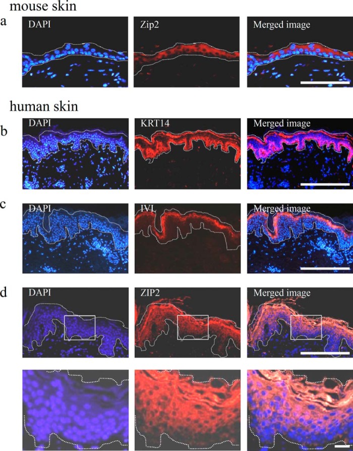
FIGURE 3.
Gene expression profiles of mouse and human skin. a, immunostaining images of mouse skin sections stained for the Zip2 antibody. As with the gene expression analysis, the expression of Zip2 was not detected in the dermis but was in the epidermis. More specifically, the expression of Zip2 was not detected in undifferentiated keratinocytes in the basal layer but was in differentiated keratinocytes in the upper layer. b–d, immunostaining images of human skin sections stained for the KRT14, IVL, and ZIP2 antibodies. The expression of KRT14 and IVL was confirmed in the basal layer and outer living layer of the epidermis, respectively. The expression of ZIP2 was not detected in undifferentiated keratinocytes in the basal layer but was in differentiated keratinocytes in the upper layer. The bottom panels in d are magnified views of the top panels. White dotted lines mark the epidermis. Scale bars = 100 μm (a), 200 μm (b–d, top panels), and 20 μm (d, bottom panels).

