FIGURE 2.
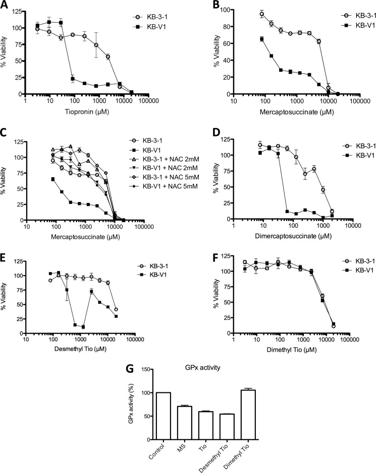
Collateral sensitivity of KB-V1 cells to inhibitors of GPx, assessed by cytotoxicity assay. A and B, dose-response curves demonstrating the effect of tiopronin (A) and mercaptosuccinate (B) on KB-3-1 and KB-V1 cells treated with tiopronin for 72 h. C, dose-response curves demonstrating the effect of (nontoxic) concentrations of N-acetylcysteine (2 and 5 mm) on the cytotoxicity of mercaptosuccinate toward KB-V1 and KB-3-1 cells. D–F, dose-response curves demonstrating the effects of dimercaptosuccinate (D), demethyl tiopronin (E), and dimethyl tiopronin (F) on KB-V1 and KB-3-1 cells. G, inhibition of bovine erythrocyte GPx activity by mercaptosuccinate, tiopronin, demethyl tiopronin, and dimethyl tiopronin (200 μm) compared with control (DMSO only) conditions.

