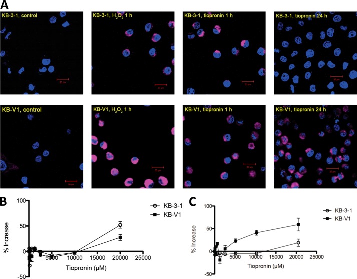
FIGURE 9.
Assessment of ROS levels in cells treated with tiopronin. A, CellROX Deep Red reagent, a dye that fluoresces when oxidized by ROS, was added to the cells and observed by confocal microscopy. KB-3-1 and KB-V1 cells were treated with tiopronin (1 mm) for 1 or 24 h. As positive controls, cells were treated with H2O2 (200 μm) for 1 h. For negative controls, cells were left untreated (i.e. dye only). Prior to imaging, all cells were incubated with Hoechst 33342 (5 μg/ml) and CellROX Deep Red reagent (5 μm). B, ROS levels generated in cells were measured by preincubating cells in 96-well plates with tiopronin for 24 h, followed by washing and incubation of cells with DHFDA (reduced form) for 30 min. The cells were then washed, and fluorescence arising from oxidation of DHFDA to fluorescein diacetate was measured. C, measurement of ROS as described above, but after the addition of dye, 1,500 μm H2O2 was applied to cells to observe the ROS levels in cells in the presence of an oxidative insult.

