FIGURE 4.
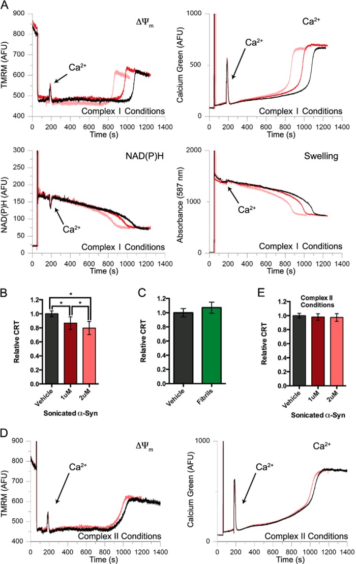
Sonicated αSyn fibrils promote Ca2+-mediated mitochondrial dysfunction in a substrate-dependent manner. A, representative traces of basic parameters of isolated liver mitochondria (ΔΨm, extramitochondrial Ca2+ fluorescence, NAD(P)H autofluorescence, and mitochondrial swelling) simultaneously measured by a multichannel fluorimeter and recorded in the presence of a single 20 μm Ca2+ addition, 5 mm glutamate and 5 mm malate as substrates, and either PBS vehicle (black traces) or 1 μm (red traces) or 2 μm (pink traces) sonicated αSyn fibrils. Spikes at 60 s result from the addition of mitochondria; arrows are used to indicate the time of the Ca2+ addition. B, the CRT of mitochondria treated with 1 and 2 μm sonicated αSyn fibrils under complex I conditions (glutamate/malate-dependent respiration) were normalized to vehicle-treated mitochondria. Sonicated αSyn dose-dependently reduced CRT under these conditions. Error bars, S.D. from at least nine independent experiments. *, p < 0.05, ANOVA followed by Tukey's multiple-comparison test. C, the relative CRT of isolated mitochondria respiring under complex I conditions and treated with non-sonicated αSyn fibrils was determined. Error bars, S.D. from five independent experiments. D, representative traces of ΔΨm (left) and extramitochondrial Ca2+ fluorescence (right) of mitochondria treated with a single aliquot of Ca2+ in the presence of succinate/rotenone (complex II-dependent respiration) and either vehicle (black traces) or 2 μm sonicated αSyn fibrils (pink traces). E, the CRT of mitochondria respiring under complex II conditions (succinate/rotenone-dependent respiration) and treated with 1 and 2 μm sonicated αSyn was compared with vehicle-treated mitochondria under identical conditions. Error bars, S.D. from at least four independent experiments.

