FIGURE 6.
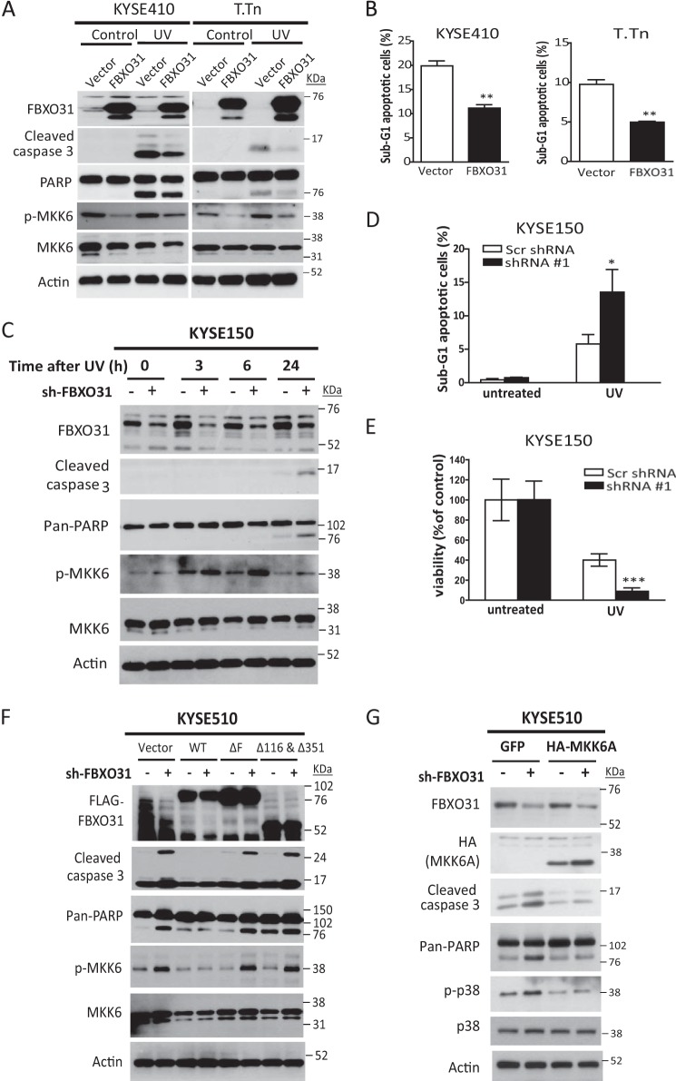
FBXO31 suppresses stress-induced cell apoptosis by modulating the MKK6-p38 pathway. A and B, serum-starved ESCC cells with stable overexpression of FBXO31 or an empty vector were treated with or without 50 J/m2 UV irradiation and incubated for 24 h. Then, cell lysates were extracted for Western blot analysis to detect changes in apoptosis markers, and endogenous phosphorylated MKK6 and MKK6 (A) or cells were harvested for flow cytometry analysis (B). The mean percentages of sub-G1 cells from three independent experiments are shown. **, p < 0.01 compared with control cells with empty vector expression. PARP, poly(ADP-ribose) polymerase. C, ESCC cells with FBXO31 knockdown were serum-starved for 24 h and then subjected to UV irradiation. Cell lysates collected at the indicated time points were immunoblotted for analysis of apoptosis and MKK6 activation. D, KYSE150 cells with the indicated shRNA expression were treated with 50 J/m2 UV irradiation for 24 h, and then cells were analyzed by flow cytometry. The mean percentages of sub-G1 cells from three independent experiments are shown. *, p < 0.05 compared with control cells expressing scrambled (Scr) shRNA. E, colony formation of UV-irradiated KYSE150 cells expressing scrambled shRNA or FBXO31-shRNA#1 cells. The bar graphs show the number of colonies relative to untreated cells. Data were calculated from three independent experiments. Error bars represent mean ± S.E. ***, p < 0.001 compared with control cells expressing Scr shRNA after UV treatment. F and G, KYSE510 cells expressing scrambled shRNA or FBXO31 shRNA#1 were transfected with the indicated plasmids and then treated with UV irradiation. Cell lysates were harvested after 6 h for Western blot analysis.

