FIGURE 7.
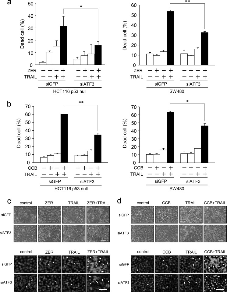
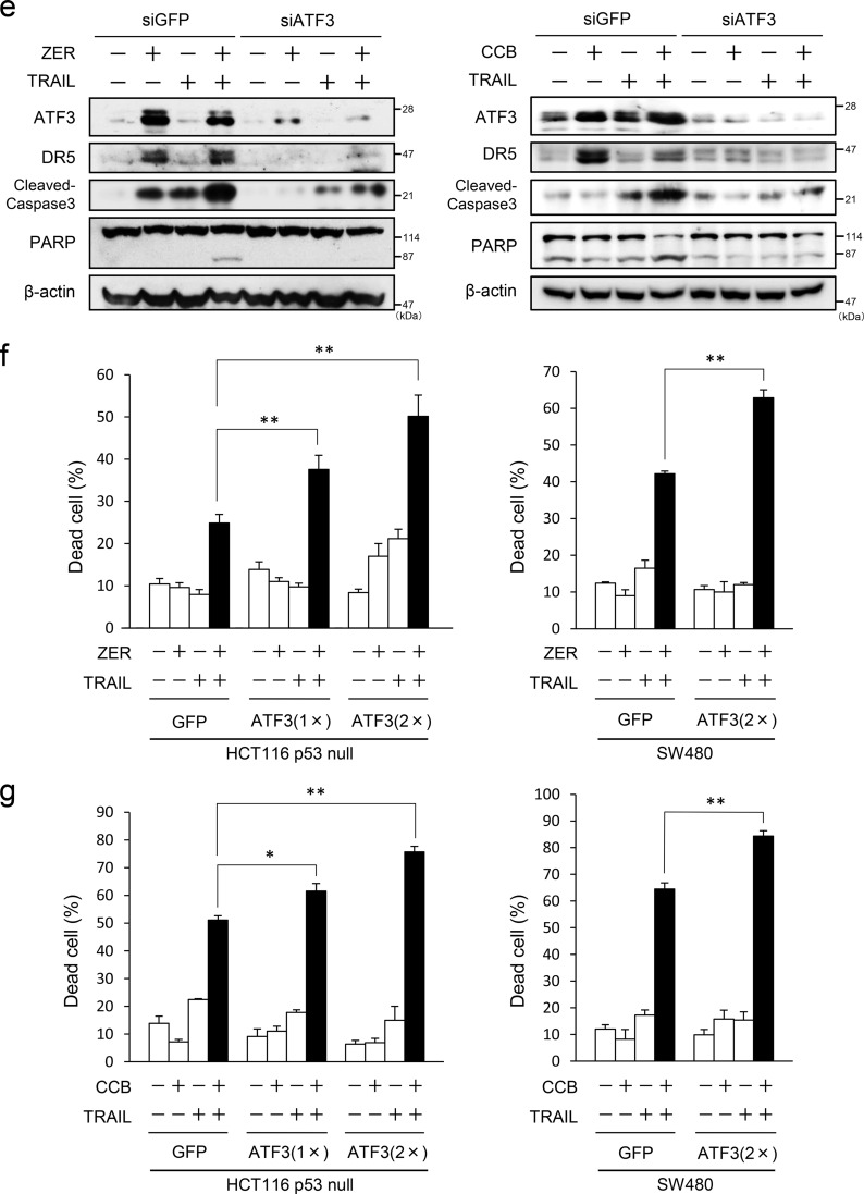
ATF3 promotes the ZER or CCB/TRAIL-induced apoptosis of p53-deficient colorectal cancer cells. a, ATF3 was knocked down in HCT116-p53null cells (left panel) or SW480 cells (right panel), and cells were treated with 30 μm ZER and/or 2.5 ng/ml TRAIL for 24 h. Cell death was measured using a trypan blue exclusion assay. The proportions of dead cells from three independent experiments are shown. b, ATF3 knockdown cells were treated with 75 μm CCB and/or 2.5 ng/ml TRAIL for 24 h. Cell death was measured as in a. HCT116-p53null cells were treated with 20 μm ZER (c) or 50 μm CCB (d) and/or 2.5 ng/ml TRAIL for 24 h. The cells were examined by phase-contrast microscopy (top panel) or fluorescence microscope after staining with DAPI (bottom panel). Arrowheads indicate cells with condensation or fragmentation of nuclei. Scale bars = 50 μm. e, whole cell extracts were prepared from cells treated as in a and analyzed by Western blotting for cleaved caspase 3, cleaved PARP, ATF3, and DR5 proteins. β-actin was used as a loading control. f, HCT116-p53null cells (left panel) or SW480 cells (right panel) stably expressing FLAG-ATF3 or GFP were treated with 30 μm ZER and/or 2.5 ng/ml TRAIL for 24 h, followed by a trypan blue exclusion assay as in a. g, ATF3 overexpressed cells as in f were treated with 75 μm CCB and/or 2.5 ng/ml TRAIL for 24 h, followed by a trypan blue exclusion assay as in a. Data are mean ± S.E. of three independent experiments. *, p < 0.05; **, p < 0.01.


