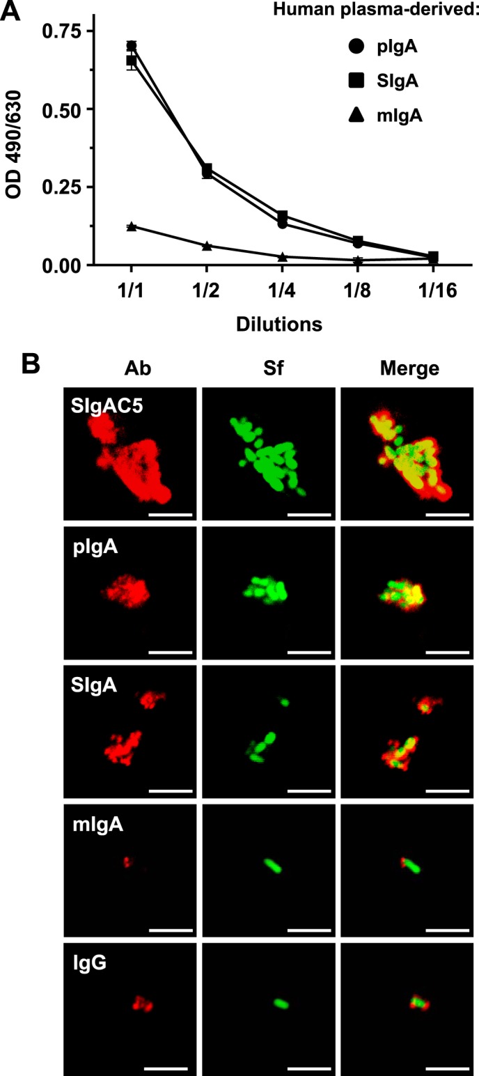
FIGURE 1.

Association of human plasma-derived IgA/SIgA with S. flexneri. A, binding of equimolar concentrations of pIgA, reconstituted SIgA, or mIgA to immobilized S. flexneri as determined by ELISA. Successive dilutions of the various molecular forms of IgA were assessed, with the 1:1 ratio corresponding to 0.61 μm of each respective Ab. Data are representative of two independent experiments performed in duplicates. B, LSCM images of immune complexes of bacteria associated with human plasma-derived pIgA, SIgA, mIgA, IgG, or anti-S. flexneri LPS-specific SIgAC5 monoclonal Ab. Bacteria constitutively expressing GFP appear in green. Bound Abs were detected by antisera directed against the α or γ chain, followed by Abs conjugated to fluorophores, yielding red signals after image processing. Images are representative of one representative field obtained from 15 observations from three independent slides per experiment. Scale bars = 10 μm.
